| Learning to Use the Microscope |
Introduction
“Micro” refers to tiny, “scope” refers to view or look at. Microscopes are tools used to enlarge images of small objects so as they can be studied. Microscopes range from a simple magnifying glass to the expensive electron microscope. The compound light microscope is the most common instrument used in education today. It is an instrument containing two lenses, which magnifies, and a variety of knobs to resolve (focus) the picture. It is a rather simple piece of equipment to understand and use. In this lab, we are going to learn the proper use and handling of the microscope.
Objectives
- Demonstrate the proper procedures used in correctly using the compound light microscope.
- Prepare and use a wet mount.
- Determine the total magnification of the microscope.
- Develop a checklist to insure the proper handling of the microscope.
Materials
- Compound microscope
- Glass slides
- Cover slips
- Eye dropper
- Beaker of water
- The letter “e” cut from newsprint
- Scissors
Procedures
Proper Handling of the Microscope
- Carry the microscope with both hands — one on the arm and the other under the base of the microscope.
- One person from each group will now go over to the microscope storage area and properly transport one microscope to your working area.
- The other person in the group will pick up a pair of scissors, newsprint, a slide, and a cover slip.
- Remove the dust cover and store it properly. Plug in the scope. Do not turn it on until told to do so.
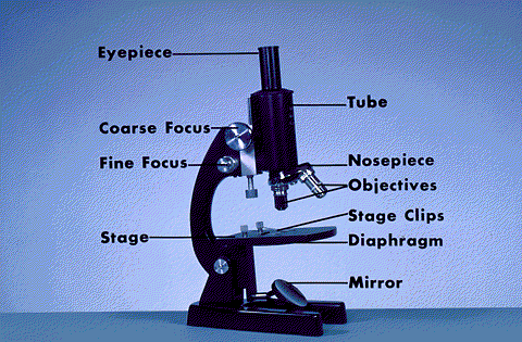
- Examine the microscope and give the function of each of the parts found below.

a. Eyepiece –
b. Body tube –
c. Objectives –
d. Stage –
f. Diaphragm –
g. Coarse adjustment –
h. Fine adjustment –
i. Base –
j. Light source –
Preparing a wet mount of the letter “e”.
- With your scissors cut out the letter “e” from the newsprint.
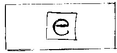
- Place it on the glass slide so as to look like (e).
- Cover it with a clean cover slip. See the figure below.

- Using your eyedropper, place a drop of water on the edge of the cover slip where it touches the glass slide. The water should be sucked under the slide if done properly.
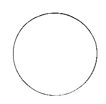
- Turn on the microscope and place the slide on the stage; making sure the “e” is facing the normal reading position (see the figure above). Using the course focus and low power, move the body tube down until the “e” can be seen clearly. Draw what you see in the space below.
- Describe the relationship between what you see through the eyepiece and what you see on the stage.
7. Offer an explanation of why this happened.
8. Looking through the eyepiece, move the slide to the upper right area of the stage.
What direction does the image move?
9. Now, move it to the lower left side of the stage. What direction does the image move?
10. Re-center the slide and change the scope to high power. You will notice the “e” is out of focus. Do Not touch the coarse focus knob, instead use the fine focus to resolve the picture.
11. Locate the diaphragm under the stage. Move it and record the changes in light intensity as you do so.
Determining Total Magnification:
1. Locate the numbers inscribed on the eyepiece and the low power objective and fill in the blanks below.
| Eyepiece magnification ______________ | (X) | Objective magnification ______________ | = | Total Magnification _____________X |
2. Do the same for the high power objective.
| Eyepiece magnification ______________ | (X) | Objective magnification ______________ | = | Total Magnification _____________X |
3. Write out the rule for determining total magnification of a compound microscope.
4. Remove the slide and clean it up. Turn off the microscope and wind up the wire so it resembles its original position. Place the low power objective in place and lower the body tube. Cover the scope with the dust cover. Place the scope back in its original space on the storage cart.
Summary:
Develop a procedure by which anyone can follow to demonstrate the proper handling of the microscope.
| BACK |

