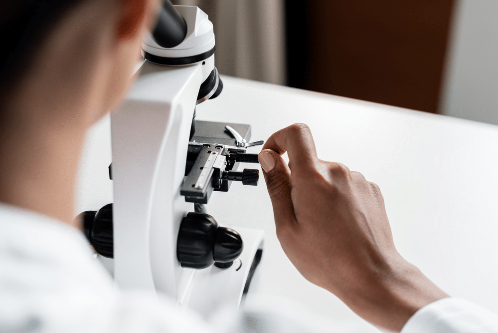
Microscopes offer a great way to discover an entire universe that lies beyond what we can see with the naked eye. From harmful bacteria to beautiful and unique crystal shapes, microscopes open an entire world for us to explore which would otherwise be impossible to learn about. To experience this vast but minuscule new world it is important to know how to prepare a microscope slide for the different materials you’ll want to examine close up.
This article serves as a simple, easy-to-follow guide on how to prepare a microscope slide. This includes a list of the materials needed to mount slides, an explanation of the different techniques of mounting slides and when to use them, what techniques to use for the best results depending upon the specimen, and which style of slides to choose for which type of observations you’ll be making. Follow this easy guide to explore what the microscopic world has to offer!
How to Prepare a Microscope Slide
Gather the Materials Needed
When considering how to prepare a microscope slide, you should first gather all the necessary materials for creating slides. As you will see later on in this instructional guide, different types of materials you wish to observe under a microscope call for different types of slide mounts. Also, the different types of observations you wish to make each have their own requirements regarding shape of the slide you should use. Regardless of what you are observing and how you will observe it, there are certain basic materials you will need. These materials include:
- Slides
- Coverslips
- Pipette (also called a dropper)
- Tweezers
- Cotton or paper towel
- Petroleum jelly
- Stains (chemical or organic)
- Fluids for wet mounting
- Samples of the material you wish to observe
Microscope slides can be made of glass or plastic, feature a flat or concave shape, and each one will have its own advantage and purpose, depending on what type of observations you will be doing. For example, plastic slides are more resilient and less likely to break, so they are safer to handle as they have no sharp edges, so they are a better choice if you’ll be preparing your microscope slides outside.
Glass slides generally have a better reflective index and are less likely to scratch, which allows for better photos to be taken of the specimens than those on plastic slides. Choosing glass or plastic slides is a personal choice, but regardless of the materials the slides are made of, the standard size of a typical microscope slide is approximately 1X3 inches and between 1mm-1.2mm thick.
Wet vs. Dry Mounts
There are two main methods of mounting microscope slides: the wet mount method and the dry mount method. The dry mount technique is simpler and is ideal for larger specimens that that are inorganic or dead matter. Feathers, pollen, hair samples, and insects are all Ideal examples for dry mounts. Thicker or opaque samples might have to be sliced thinly to allow light to pass through the specimen which will help you see things better under the microscope. Because these samples are lifeless, these slides rarely expire and can be preserved for longer periods of time.
Wet mounts are more complex and require more attention, so keep this in mind when planning how to prepare a microscope slide. Generally used for observing organisms that live in water and other liquids, such oils, glycerin, and brine, wet mounts are also useful for when the material itself is a fluid, such as observing blood. Anything that doesn’t require the addition of water to be observed under a microscope needs to be prepared on a wet mount.
It is also important to note that using a wet mount technique has its limitations concerning living organisms. Because wet mount slides will ultimately dehydrate the living organisms within the slide, those organisms have a limited lifespan while on the slide, and therefore there is a limited shelf life for the slide itself.
For example, certain organisms, such as protozoa, offer us a very limited window of observation, as they can only survive in a wet mount slide for approximately 30 minutes if the slide is allowed to dehydrate. A way to slow this process down and have more observation time in this situation would be to seal the edges of the slide with petroleum jelly. This way, the liquid will remain in the slide longer and the life of the slide will be extended for a few days.
Another issue concerning wet mount slides involves specimens that are too large to allow the coverslip to be placed comfortably on top and rest flatly on top. Here, you might place ground pieces of glass from a spare coverslip to encase the specimen to provide some extra space for the specimen to be secured. You may also place a small cotton strand around the edge to perform the same function and corral the specimen in place. This is also a great technique to use when live specimens are quick moving, as this will limit their movement and slow them down, giving you a better observation experience.
Smears, Squash, and Stains–How and When to Use Each
Knowing how to prepare a microscope slide properly also involves applying the proper technique, as different techniques are used depending on the material being observed. Depending upon which type of material you will be looking at under your microscope, you should use the right technique to get the best results. Using these three techniques under the right circumstances shows you are certain in how to prepare a microscope slide properly.
Smear Slides
Smear slides are fairly straightforward and create microscope slides that look exactly as the name suggests: a thin smear of material across the clear slide. This method is primarily used for blood samples or samples that are fluid in nature. This is done by using a pipette (or dropper) to place a drop of the material onto the slide. Using a second slide to smear the material across the first, you can create a very thin coating that allows for clear observation. This slide creation technique allows the specimen to dehydrate at a moderate pace.
Squash Slides
Squash slides are a way to prepare soft material for observation. Drop the fluid of choice onto the slide and press down slightly as to flatten the sample and squeeze the liquid from it without breaking the slide or coverslip. Use a tissue to absorb the excess liquid. This wet mounting technique is ideal for tissue or sponge samples.
Stain Solutions
Stain applications are a great way to distinguish between living and non-living cells in your specimen sample. This technique is primarily done in the biological science labs to help scientists identify diseases, especially different bacteria, and examine the minute characteristics of cells more closely.
Depending on what exactly you are trying to identify, there are several types of stains you can use, but the most common is iodine. Prepare the wet mount as you would with any other fluid, in this case using the staining solution, place the coverslip on the edge of the slide, and slowly pull the stained liquid sample across the slide. Use a paper towel to absorb the excess liquid.
Flat vs. Concave Slides—Which to Choose?
When first learning how to prepare a microscope slide, it is important to consider what type of material you will be observing. It is equally important to consider what type of observation will be best based on the consistency of the material of your sample. This is where you will decide whether you want to preserve your slide and keep it for further use, or if that is not possible, perhaps it is more practical to not use a coverslip for your wet mount. But how can you made observations under your microscope without a coverslip?
This is made possible through the use of a concave style slide. Also known as a depression slide or a well slide, this microscope slide is shaped so it can hold a drop of liquid in an indentation without the use of a cover. As expected, this option is considerably more expensive, but will allow you to observe a live organism and preserve it for future observation as flat wet mounts will shorten the life span of the specimen considerably. Concave slides also allow for free movement of specimens within the drop of water or fluid present.
Conclusion
Microscopes can lift the veil on a whole new world for you, your friends, and family, especially know that you know the various aspects about preparing microscope slides. Knowing how to prepare a microscope slide properly lets you to observe a variety of materials, witness what changes occur over time, compare specimens, and potentially preserve those specimens indefinitely! Learning how to prepare a microscope slide properly offers many benefits, and we hope this quick guide has given you the confidence you need to prepare slides of your own while you’re out in the field or in your home laboratory.

